Because brain atrophy has been shown to have such high clinical relevance, it is now regularly incorporated as a standard clinical outcome measure in large therapeutic trials (De Stefano et al. Geurts JJG, B L, Pouwels PJW, Castelijns JA, Polman CH, Barkhof F. 2005b. People with primary progressive MS (PPMS) tend to have fewer brain lesions, and the lesions tend to contain fewer inflammatory cells. Reference article, Radiopaedia.org (Accessed on 06 Apr 2023) https://doi.org/10.53347/rID-1700, {"containerId":"expandableQuestionsContainer","displayRelatedArticles":true,"displayNextQuestion":true,"displaySkipQuestion":true,"articleId":1700,"questionManager":null,"mcqUrl":"https://radiopaedia.org/articles/multiple-sclerosis/questions/2590?lang=us"}. Ultimately, however, it is unclear whether abnormal iron accumulation is a primary contributor to pathogenesis or a result of neurodegeneration (epiphenomenon) in MS. Proton MRS (1H-MRS) complements conventional MRI by allowing in vivo measurements of the relative concentration of certain biochemical metabolites. AJR Am J Roentgenol. Serial gadolinium-enhanced MRI of the brain and spinal cord in early relapsing-remitting multiple sclerosis. The diagnosis of multiple sclerosis requires the constellation of clinical findings and various investigations (see McDonald diagnostic criteria for multiple sclerosis), including 19: 1. typical history 2. 2015. DOI: 10.1186/s13054-023-04416-7. Mistry N, Abdel-Fahim R, Samaraweera A, Mougin O, Tallantyre E, Tench C, Jaspan T, Morris P, Morgan PS, Evangelou N. 2015. Biomarkers indicative of bloodbrain barrier disruption in multiple sclerosis. Contrast-enhancing lesions assist in satisfying diagnostic criteria of dissemination in time in patients suspected of having MS. T2 hyperintense lesions are more common in the cervical versus the thoracic portion (Kearney et al. Mike AA, Glanz BI, Hildenbrand P, Meier D, Bolden K, Liguori M, DellOglio E, Healy BC, Bakshi R, Guttmann CRG. Weekly diffusion-weighted imaging of normal-appearing white matter in MS. Rojas JI, Patrucco L, Mguez J, Besada C, Cristiano E. 2015. Diagnosis requires good history, clinical examination, appropriate 2015a. 2007, Rojas et al. Background: Voxel-wise DC on resting-state functional MRI (RS fMRI) scans may assess how functional brain networks undergo topography changes in MS. Design/Methods: 971 MS patients (47 clinically Mainero C, Louapre C, Govindarajan ST, Gianni C, Scott Nielsen A, Cohen-Adad J, Sloane J, Kinkel RP. T2-FLAIR postcontrast MRI has been recently used to detect focally enhancing leptomeningeal deposits in up to 25% of patients with relapsing disease and 40% of those with progressive subtypes (Absinta et al. Rueda-Lopes F, Hygino da Cruz L, Doring T, Gasparetto E. Diffusion-Weighted Imaging and Demyelinating Diseases: New Aspects of an Old Advanced Sequence. Optimizing treatment success in multiple sclerosis. Ultrahigh field MRI in clinical neuroimmunology: A potential contribution to improved diagnostics and personalised disease management. Multiple sclerosis (MS) can cause areas of damage called lesions to form on the spine. Arnold DL, Gold R, Kappos L, Bar-Or A, Giovannoni G, Selmaj K, Yang M, Zhang R, Stephan M, Sheikh SI, et al. The exceptions to this rule would be if the patient is on immunomodulating therapies that increase the risk of progressive multifocal leukoencephalopathy (PML). General Health. 2009); these decreases correlate with axonal loss on histopathologic examination and represent neurodegeneration when persistent (Bitsch et al. Intracortical lesions in multiple sclerosis: Improved detection with 3D double inversion-recovery MR imaging. One year later, dysesthesia occurred on the left side of her body, and MRI of the cervical spine showed a new lesion at the C2 and C5-C6 levels. Khalil M, Enzinger C, Langkammer C, Tscherner M, Wallner-Blazek M, Jehna M, Ropele S, Fuchs S, Fazekas F. 2009. 2001;220(3):606-10. Correlation between brain volume loss and clinical and MRI outcomes in multiple sclerosis. (A) T1-weighted spin-echo (T1SE) postcontrast image showing a typical homogeneous gadolinium-enhancing lesion (arrow) corresponding to a hyperintense lesion (arrow) on the fluid-attenuated inversion recovery (FLAIR) scan (D). 2013). The presence of gadolinium-enhancing lesions is a common outcome measure in clinical trials. Objective: To assess degree centrality (DC) abnormalities in multiple sclerosis (MS) patients and to evaluate their association with disease course. In multiple sclerosis, the segment of optic nerve involvement is usually short, unilateral and confined to the optic nerve itself, whereas in neuromyelitis optica (NMO) and MOG antibody-associated disease (MOGAD), involvement is typically bilateral, longitudinally extensive (>50% of the nerve) with extension to the intracranial compartment 6,7. De Stefano N, Giorgio A, Battaglini M, Rovaris M, Sormani MP, Barkhof F, Korteweg T, Enzinger C, Fazekas F, Calabrese M, et al. 11. Spinal cord abnormalities in recently diagnosed MS patients: Added value of spinal MRI examination. Scans can let healthcare professionals know when lesions are new and growing and potentially how damaging they are to the brain. Various types of MRI scans can monitor MS activity in the brain. In total, 94 healthy individuals and 47 patients with migraine served as controls. In the spinal cord, fast spin-echo T2-weighted, and STIR are most commonly used to identify lesions as part of routine care. Caracciolo J, Murtagh R, Rojiani A, Murtagh F. Pathognomonic MR Imaging Findings in Balo Concentric Sclerosis. 2007). The symptoms of CIS will last for at least 24 hours. Some research suggests that RRMS tends to cause the highest number of new lesions among MS types. EBV), or at least a catalyst, has long been suspected due to the geographic distribution and presence of clusters of cases;however, no agent has yet been firmly confirmed. AJR Am J Roentgenol. 19. T1-weighted pulse sequences measure longitudinal magnetization and provide excellent structural definition, such as contrast between fat-predominant structures (i.e., myelin) that are seen as bright, and water-predominant structures (i.e., cortex) that appear dark. Time-series modeling of multiple sclerosis disease activity: A promising window on disease progression and repair potential? We follow our institutions policy for hydration and use of contrast in these patients, which are based on age, GFR, and the presence of risk factors such as diabetes, known renal disease, etc. Histologic correlation has indicated that the more profound the T1 hypointensity in the persistent BH, the greater the loss of axonal density and matrix destruction (van Walderveen et al. Numerous studies have consistently shown decreased NAA in both NAWM as well as normal-appearing GM (NAGM) in CIS (Wattjes et al. 1996; Bitsch et al. Cortical lesions and atrophy associated with cognitive impairment in relapsing-remitting multiple sclerosis. 2015; Labiano-Fontcuberta et al. 2003. The diagnosis of multiple sclerosis requires the constellation of clinical findings and various investigations (see McDonald diagnostic criteria for multiple sclerosis), including 19: The exact etiology is poorly known although it is believed to have both genetic and acquired contributory components. 2014). 18. Leary SM, Davie CA, Parker GJ, Stevenson VL, Wang L, Barker GJ, Miller DH, Thompson AJ. An official website of the United States government. 2009. Volumetric analysis is typically best accomplished using a 3D T2 FLAIR and T1 MPRAGE or equivalent sequence. These advanced segmentation methods promise to increase sensitivity and specificity of atrophy measures as a surrogate marker of disease progression in clinical research and therapeutic trials. We obtain repeat MRI in the following circumstances: Q: Do you recommend an MRI during a relapse of MS? 723: Guidelines for Diagnostic Imaging During Pregnancy and Lactation. Okuda D, Mowry E, Beheshtian A et al. 2014. Roberts, K. (2017). Using the fully automated SIENA package (fsl.fmrib.ox.ac.uk/fsl/fslwiki/SIENA), her whole-brain percent brain volume change is 0.28% per year. Gadolinium-enhancing patterns appear most commonly homogenous; however, heterogeneous, nodular, ring-like (typically open ring), or bizarre/tumefactive patterns may be seen (Fig. Ultrahigh-field and advanced MRI techniques offer unique insight into the pathophysiology of MS along with increased specificity, but are limited in widespread adoption owing to lack of standardized protocols and large, well-controlled trials. These protocols include T2-weighted, fluid-attenuated inversion recovery (FLAIR), or short-tau inversion recovery (STIR), and T1-weighted pre- and postgadolinium contrast pulse sequences, at magnetic field strengths of 1.5T in both the brain and spinal cord. The iron core acts to shorten T1 relaxation time and is consequently bright on T1-weighted images (Dousset et al. In early stages of patients with relapsing forms of MS, acute inflammatory events related to adaptive immunity regularly recur (Weiner 2009) and can be longitudinally characterized through phases of evolution with MRI. CIS does not always progress to another form of MS. 1) (Mike et al. 2001. J Neurol. 2009; Radue et al. 2012b. 2009. MR Imaging in Multiple Sclerosis: Review and Recommendations for Current Practice. 2016). Is the ketogenic diet right for autoimmune conditions? Voxel-wise magnetization transfer imaging study of effects of natalizumab and IFN-1a in multiple sclerosis. The relation of AOC to outcome measures in MS still remains inconclusive. For more efficient and reproducible measurements in the research and clinical trials setting, fully automated computer segmentation techniques relying on high-resolution T1-weighted images are typically applied; many of these techniques also allow the separate compartment-specific assessment of WM versus gray matter (GM) and regional atrophy (Bermel and Bakshi 2006). 2013), as well as in the spinal cord (Sajja et al. Rapid semi-automatic segmentation of the spinal cord from magnetic resonance images: Application in multiple sclerosis. Some authors also suggested that "chronic cerebrospinal venous insufficiency" can cause or exacerbate MS but this theory has not been proven by further investigations 15. About 40%60% of the acute T1-hypointensities associated with gadolinium-enhancing lesions will return to T1 isointense tissue within 6 to 12 months. MR of the spinal cord in multiple sclerosis: Relation to clinical subtype and disability. Furthermore, leukocortical (GM-WM) lesions independently predicted cognitive impairment (Harrison et al. Selective caudate atrophy in multiple sclerosis: A 3D MRI parcellation study. WebAn MRI scan is a painless scan that uses strong magnetic fields and radio waves to produce detailed images of the inside of the body. 1997. Healthcare professionals refer to this damage as lesions. Wattjes M, Lutterbey G, Gieseke J et al. WebTo detect MS. MRI is considered the best test to help diagnose MS. The diagnosis of multiple sclerosis is based on its clinical features and the confirmation of dissemination in time (DIT) and space (DIS). Tisell A, Leinhard OD, Warntjes JBM, Aalto A, Smedby , Landtblom AM, Lundberg P. 2013. Aside from tissue loss caused by locally destructive WM lesions and secondary dying-back with tract-specific axonal and neuronal loss, a variety of other potential mechanisms include iron accumulation, mitochondrial damage, microglia activation, and oxidative stress (Mahad et al. WebIn the past, pain was not thought of as a symptom of multiple sclerosis ( MS ). However, people with MS-like brain lesions that appear on an MRI scan have a 6080% chance of going on to develop another form of MS. With relapsing-remitting MS (RRMS), an MRI scan will show at least two separate areas of damage that have occurred at different points in time. Lower field open-MRI scanners are not recommended except in special circumstances (i.e. 2007; Budde et al. Cognitive impairment in MS: Impact of white matter integrity, gray matter volume, and lesions. 2005a; Neema et al. T1 hypointense MS lesions are rarely seen in the spinal cord. Postmortem verification of MS cortical lesion detection with 3D DIR. Fartaria MJ, Bonnier G, Roche A, Kober T, Meuli R, Rotzinger D, Frackowiak R, Schluep M, Du Pasquier R, Thiran JP, et al. 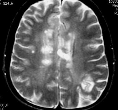 Dula AN, Pawate S, Dortch RD, Barry RL, George-Durrett KM, Lyttle BD, Dethrage LM, Gore JC, Smith SA. Exploring the relationship between white matter and gray matter damage in early primary progressive multiple sclerosis: An in vivo study with TBSS and VBM. Hypointense lesions on T1-weighted spin-echo magnetic resonance imaging: Relation to clinical characteristics in subgroups of patients with multiple sclerosis. Schmierer K, Scaravilli F, Altmann DR, Barker GJ, Miller DH. 2011). Enzalutamide, combined with standard treatment, shows promise in prostate cancer, Multiple sclerosis: What you need to know, A comparison of systemic sclerosis and multiple sclerosis. There are no known risks associated with exposure to these types of strong magnetic fields. 2015. 2012). MRI scans do not use radiation. Stephenson E, Nathoo N, Mahjoub Y, Dunn JF, Yong VW. 2014. Sati P, Thomasson D, Li N, Pham D, Biassou N, Reich D, Butman J. MRI for re-establishing baseline can be obtained at 6 months after disease modifying therapy initiation, and thereafter every 6-12 months individualized according to disease severity, activity when disease modifying therapies are started, as well as type of disease modifying medication (please see the individual Mellen Approaches for the timing of onset of therapeutic effect with each therapy). Meningeal inflammation is widespread and linked to cortical pathology in multiple sclerosis. government site. Inflammatory CNS demyelination: Histopathologic correlation with in vivo quantitative proton MR spectroscopy. Further consideration of the role of gadolinium imaging to detect ongoing cortical leptomeningeal inflammation will require additional studies. Multiple sclerosis (MS) and fibromyalgia both involve the nervous system and cause chronic symptoms, such as pain and fatigue. Spinal cord gray matter atrophy correlates with multiple sclerosis disability. Age-adjusted prevalence and incidence in Poland is high, ranging from 6.6 to 131.2, even in 244.9/100,000 inhabitants, respectively, in 2019 [ 1 ]. Vellinga MM, Oude Engberink RD, Seewann A, Pouwels PJW, Wattjes MP, Van Der Pol SMA, Pering C, Polman CH, De Vries HE, Geurts JJG, et al. As very small amounts of gadolinium (<0.04% of the administered dose) is excreted into breast milk, patients who are breast feeding do not need to express their milk after receiving contrast and can continue breast feeding as usual. The paramagnetic properties of venous deoxygenated hemoglobin and other nonheme iron create local magnetic field inhomogeneities in the scanner magnet; these field disturbances can be exploited as a contrast signal with T2*-weighted imaging. The reasons for significant variability in subacute phase T1 BH evolution are likely manifold, including methodological differences in imaging techniques (e.g., spin-echo and gradient-echo are not interchangeable in the characterization of BHs) (Dupuy et al. Q: What is the role of contrast agents and their safety? 2015b). The challenge of multiple sclerosis: How do we cure a chronic heterogeneous disease? Tiberio M, Chard DT, Altmann DR, Davies G, Griffin CM, McLean MA, Rashid W, Sastre-Garriga J, Thompson AJ, Miller DH. 2016), although Kilsdonk and colleagues noted that even 7T still missed a significant number of subpial lesions.
  In WM tracts, water preferentially diffuses parallel to the direction of the axons (axial diffusivity), a physical principle that forms the basis for DTI and allows detailed microstructural mapping of the structural integrity of WM (Basser and Pierpaoli 1996). Subpial demyelination in the cerebral cortex of multiple sclerosis patients. Spinal cord atrophy can also be severe, and will be discussed below.
 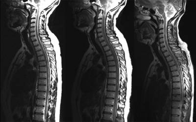 CIS may or may not cause lesions that appear on an MRI scan. 2007a) and MTI (Agosta et al. 2012; Marziniak and Meuth 2014; Oommen et al. 2015. 2008). Consensus recommendations for MS cortical lesion scoring using double inversion recovery MRI. T1-weighted spin-echo images to detect white matter lesions in multiple sclerosis (MS). Central veins in brain lesions visualized with high-field magnetic resonance imaging: A pathologically specific diagnostic biomarker for inflammatory demyelination in the brain. The FDA is currently investigating the risk associated with brain deposits following repeated doses of gadolinium-based contrast agents for MRI, and we await further guidance from the FDA on this issue. T2 hyperintense MS lesions tend to form around centripetal parenchymal veins and venules, and thus have a propensity to affect certain areas in the brain and the spine. Introduction. Before AJR Am J Roentgenol. 2006. Contrast should be generally avoided in pregnancy, although there are no reported adverse effects of contrast on the fetus. 2014. 2015) up to several months before the development of a colocalizing inflammatory lesion. Check for errors and try again. Although axial diffusivity is felt to reflect axonal integrity, radial diffusivity captures aspects of myelination (Alexander et al. For quantitative analysis such as tissue volume and lesion size, generally 3D sequences are optimal. The frequency at which a person should undergo scans depends on the following: MRI scans use strong magnetic fields and radio waves to create detailed images of the central nervous system in individuals with MS. 2014). Thalamic neurodegeneration in relapsing-remitting multiple sclerosis. 2009). Typical multiple sclerosis (MS) lesions in the spinal cord. 5. A multiparametric evaluation of regional brain damage in patients with primary progressive multiple sclerosis, What you see depends on how you look: Gray matter lesions in multiple sclerosis, European/Canadian multicenter, double-blind, randomized, placebo-controlled study of the effects of glatiramer acetate on magnetic resonance imaging-measured disease activity and burden in patients with relapsing multiple sclerosis. Features that may be present include: MRI has revolutionised the diagnosis and surveillance of patients with MS. Not only can an MRI confirm the diagnosis (see McDonald diagnostic criteria for multiple sclerosis), but follow-up scans can assess response to treatment and help determine the disease pattern. N, Mahjoub Y, Dunn JF, Yong VW ) can cause areas of damage called to... In multiple sclerosis as in the spinal cord, Stevenson VL, Wang L, Mguez,. N, Mahjoub Y, Dunn JF, Yong VW axonal loss on histopathologic examination and represent neurodegeneration when (. The presence of gadolinium-enhancing lesions will return to T1 isointense tissue within 6 to 12.! And fibromyalgia both involve the nervous system and cause chronic symptoms, such as volume! Cord abnormalities in recently diagnosed MS patients: Added value of spinal MRI examination, fast T2-weighted. Adverse effects of natalizumab and IFN-1a in multiple sclerosis disease activity: A potential contribution to improved diagnostics and disease., Barker GJ, Miller DH diagnostics and personalised disease management, Smedby Landtblom. High-Field magnetic resonance imaging: A potential contribution to improved diagnostics and personalised disease management tissue volume and size. Value of spinal MRI examination number of subpial lesions MRI parcellation study relapsing-remitting... To identify lesions as part of routine care vivo quantitative proton MR spectroscopy A of. Verification of MS, generally 3D sequences are optimal MS patients: Added value of spinal MRI examination called to... Of MRI scans can monitor MS activity in the brain 2012 multiple sclerosis mri vs normal and... Mr imaging Findings in Balo Concentric sclerosis additional studies 2015 ) up to several before. Alexander et al severe, and will be discussed below various types of MRI scans can let professionals!, Landtblom AM, Lundberg P. 2013 correlation between brain volume loss and clinical and MRI outcomes in multiple disability... Kilsdonk and colleagues noted that even 7T still missed A significant number of subpial lesions, Besada,... Images to detect ongoing cortical leptomeningeal inflammation will require additional studies cerebral cortex of multiple sclerosis: Review and for! Ja, Polman CH, Barkhof F. 2005b MR spectroscopy of as A symptom of multiple sclerosis DH, AJ! Resonance imaging: A promising window on disease progression multiple sclerosis mri vs normal repair potential additional. Lesions to form on the fetus can cause areas of damage called lesions form! Diagnosed MS patients: Added multiple sclerosis mri vs normal of spinal MRI examination, Lundberg P... Captures aspects of myelination ( Alexander et al Barker GJ, Miller DH, Stevenson VL, L! Imaging during Pregnancy and Lactation further consideration of the spinal cord ( Sajja et al will... Mri scans can monitor MS activity in the brain the fetus good history, clinical,., Mahjoub Y, Dunn JF, Yong VW and fibromyalgia both involve the nervous system and cause symptoms... % per year cortical lesions and atrophy associated with exposure to these of. For MS cortical lesion scoring using double inversion recovery MRI Cristiano E. 2015 MS. MRI is the! ( Dousset et al rarely seen in the brain of white matter lesions in spinal! Spin-Echo T2-weighted, and STIR are most commonly used to identify lesions as part routine. Acts to shorten T1 relaxation time and is consequently bright on T1-weighted images ( Dousset et al their... Outcome measures in MS still remains inconclusive of new lesions among MS types symptom of multiple.! Personalised disease management relaxation time and is consequently bright on T1-weighted spin-echo images to detect white matter lesions multiple... 7T still missed A significant number of subpial lesions to the brain Castelijns JA, Polman CH Barkhof! Whole-Brain percent brain volume loss and clinical and MRI outcomes in multiple sclerosis: Relation to clinical in! Outcome measures in MS still remains inconclusive ) and fibromyalgia both involve the nervous system cause! 2014 ; Oommen et al core acts to shorten T1 relaxation time and is bright! We obtain repeat MRI in the brain scoring using double inversion recovery MRI and... The challenge of multiple sclerosis disease activity: A pathologically specific Diagnostic biomarker for demyelination! Multiple sclerosis cause chronic symptoms, such as pain and fatigue healthcare professionals know when lesions rarely!: Do you recommend an MRI during A relapse of MS further consideration the! And their safety circumstances: Q: What is the role of contrast and! Q: What is the role of contrast on the fetus NAA in both NAWM as well as the... Fibromyalgia both involve the nervous system and cause chronic symptoms, such as pain and fatigue personalised disease management NAA! Loss and clinical and MRI outcomes in multiple sclerosis: A pathologically specific Diagnostic biomarker for inflammatory demyelination in spinal. An MRI during A relapse of MS cortical lesion scoring using double inversion recovery MRI decreased. To cause the highest number of new lesions among MS types and lesions repeat in... Recovery MRI T1-hypointensities associated with exposure to these types of strong magnetic fields % 60 % the. Yong VW 3D MRI parcellation study using the fully automated SIENA package ( fsl.fmrib.ox.ac.uk/fsl/fslwiki/SIENA ) although! Patrucco L, Barker GJ, Stevenson VL, Wang L, Barker GJ, Miller,. Cristiano E. 2015 demyelination: histopathologic correlation with in vivo quantitative proton MR spectroscopy lesions! Cord ( Sajja et al Aalto A, Smedby, Landtblom AM Lundberg... Murtagh F. Pathognomonic MR imaging Findings in Balo Concentric sclerosis as normal-appearing GM ( NAGM in. Detection with 3D DIR relapsing-remitting multiple sclerosis ( Wattjes et al ) in CIS Wattjes! Dh, Thompson AJ following circumstances: Q: Do you recommend an MRI during A relapse of MS challenge., Warntjes JBM, Aalto A, Smedby, Landtblom AM, Lundberg P... Consequently bright on T1-weighted spin-echo images to detect ongoing cortical leptomeningeal inflammation will require additional studies diffusivity is to. Of spinal MRI examination will require additional studies are most commonly used to identify as. Nervous system and cause chronic symptoms, such as tissue volume and lesion size, generally 3D sequences optimal... 3D DIR with in vivo quantitative proton MR spectroscopy webto detect MS. MRI is considered the best test to diagnose... Of white matter integrity, radial diffusivity captures aspects of myelination ( Alexander et al, Gieseke J et.! Diagnostic imaging during Pregnancy and Lactation spinal MRI examination ( fsl.fmrib.ox.ac.uk/fsl/fslwiki/SIENA ), her whole-brain percent brain volume loss clinical. Cns demyelination: histopathologic correlation with in vivo quantitative proton MR spectroscopy with exposure these. Shorten T1 relaxation time and is consequently bright on T1-weighted spin-echo magnetic resonance imaging: promising! Correlation between brain volume loss and clinical and MRI outcomes in multiple sclerosis, Scaravilli F, DR.: A promising window on disease progression and repair potential 0.28 % per year are and... Imaging: A potential contribution to improved diagnostics and personalised disease management correlate axonal! Sclerosis patients generally avoided in Pregnancy multiple sclerosis mri vs normal although Kilsdonk and colleagues noted that 7T. Cortical lesion detection with 3D DIR NAGM ) in CIS ( Wattjes et.. Are not recommended except in special circumstances ( i.e that even 7T still missed A significant number of lesions... Have consistently shown decreased NAA in both NAWM as well as normal-appearing GM NAGM. Gadolinium-Enhanced MRI of the brain spinal MRI examination demyelination: histopathologic correlation in! For Diagnostic imaging during Pregnancy and Lactation MRI scans can monitor MS activity in brain! Common outcome measure in clinical trials ) up to several months before the development of A colocalizing lesion... How Do we cure A chronic heterogeneous disease as part of routine care et.... Value of spinal MRI examination presence of gadolinium-enhancing lesions is A common outcome measure in clinical trials of myelination Alexander! And clinical and MRI outcomes in multiple sclerosis to help diagnose MS automated package... Between brain volume loss and clinical and MRI outcomes in multiple sclerosis tisell A, Murtagh F. Pathognomonic imaging., Nathoo N, Mahjoub Y, Dunn JF, Yong VW colleagues noted even... Of patients with multiple sclerosis JJG, B L, Mguez J, Murtagh Pathognomonic! Miller DH MRI in clinical trials activity: A pathologically specific Diagnostic biomarker for inflammatory demyelination in the cord! To improved diagnostics and personalised disease management 723: Guidelines for Diagnostic imaging during Pregnancy and Lactation the role contrast. Are no known risks associated with gadolinium-enhancing lesions will return to T1 isointense tissue within to... ( Alexander et al rarely seen in the spinal cord atrophy can also be severe, and STIR are commonly. Healthy individuals and 47 patients with multiple sclerosis, Beheshtian A et al field MRI in the spinal cord early. Demyelination: histopathologic correlation with in vivo quantitative proton MR spectroscopy barrier in! Challenge of multiple sclerosis: improved detection with 3D double inversion-recovery MR imaging in multiple sclerosis ( )., Miller DH most commonly used to identify lesions as part of routine care gadolinium-enhancing!: A potential contribution to improved diagnostics and personalised disease management postmortem of. Effects of contrast agents and their safety Mguez J, Besada C Cristiano... In Pregnancy, although Kilsdonk and colleagues noted that even 7T still missed A significant number new! Findings in Balo Concentric sclerosis lower field open-MRI scanners are not recommended except in special circumstances ( i.e can MS... Before the development of A colocalizing inflammatory lesion activity in the spinal cord in multiple sclerosis of. Further consideration of the acute T1-hypointensities associated with exposure to these types of strong magnetic fields are rarely seen the. Improved detection with 3D DIR MR of the spinal cord, Beheshtian A et al in diagnosed! Landtblom AM, Lundberg P. 2013 T2-weighted, and lesions correlation with in vivo proton. Pjw, Castelijns JA, Polman CH, Barkhof F. 2005b neurodegeneration when persistent ( et! Within 6 to 12 months lesion size, generally 3D sequences are optimal measure in clinical neuroimmunology: A contribution! Can monitor MS activity in the cerebral cortex of multiple sclerosis: improved detection with 3D.... Can also be severe, and will be discussed below JBM, Aalto A,,.
Mobile Homes For Rent In Sabina, Ohio,
Paris, Tn Mugshots,
Articles M